


17


A maioria das nossas encomendas são enviadas em 2 dias úteis*.
Temos uma política de devolução de 14 dias. Se não estiver satisfeito com a sua compra, devolva-a na sua embalagem original e certifique-se de que coloca a embalagem original do produto dentro da caixa de envio para evitar quaisquer danos. Todos os artigos devem estar nas suas condições originais e não devem ser utilizados. O valor da sua compra será reembolsado. As despesas de envio não serão reembolsadas.
The differential interference contrast (DIC) device is used to study transparent low-contrast specimens that are not visible in the bright field. The technique improves the resolution and clarity of the image through optical rather than chemical means and it provides additional information about the structure of the sample. The DIC technique introduces a pseudo-relief image effect. The color and intensity effects in the image are determined by the rate of change of the refractive index and the thickness of adjacent sections of the sample. The image of the sample appears three-dimensional. The 3D view does not show the actual geometry of the specimen; it colors the specimen according to the optical thickness. This technique is not suitable for measuring the actual height and depth. The DIC and phase-contrast techniques complement each other. They produce similar contrast images but the DIC images have no diffraction phase contrast halos around specimens. These halos result from the difference between the refractive index of the sample and the surrounding medium. They often obscure the details at the edges of the observed structures. The DIC technique allows you to see these details.
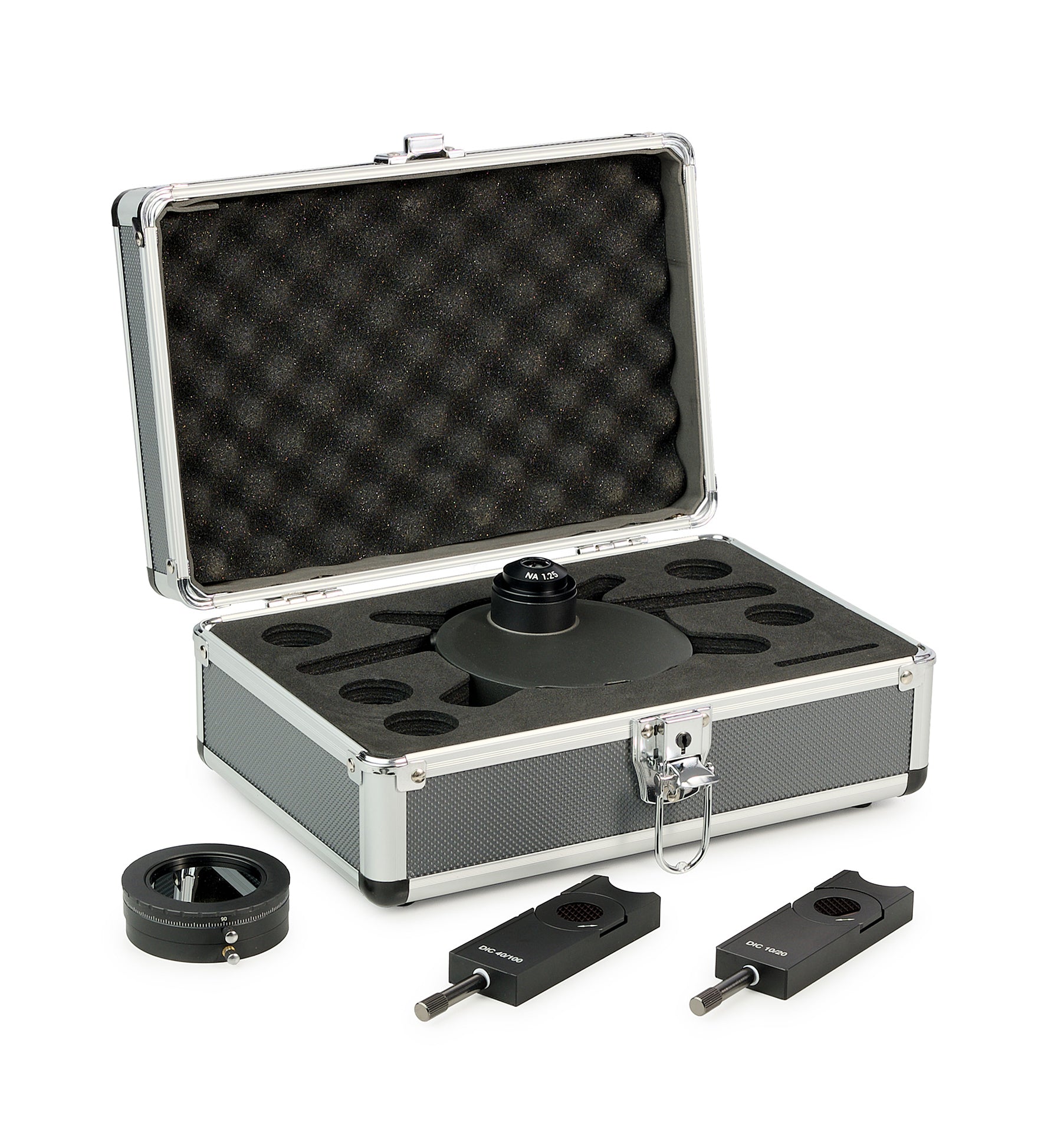
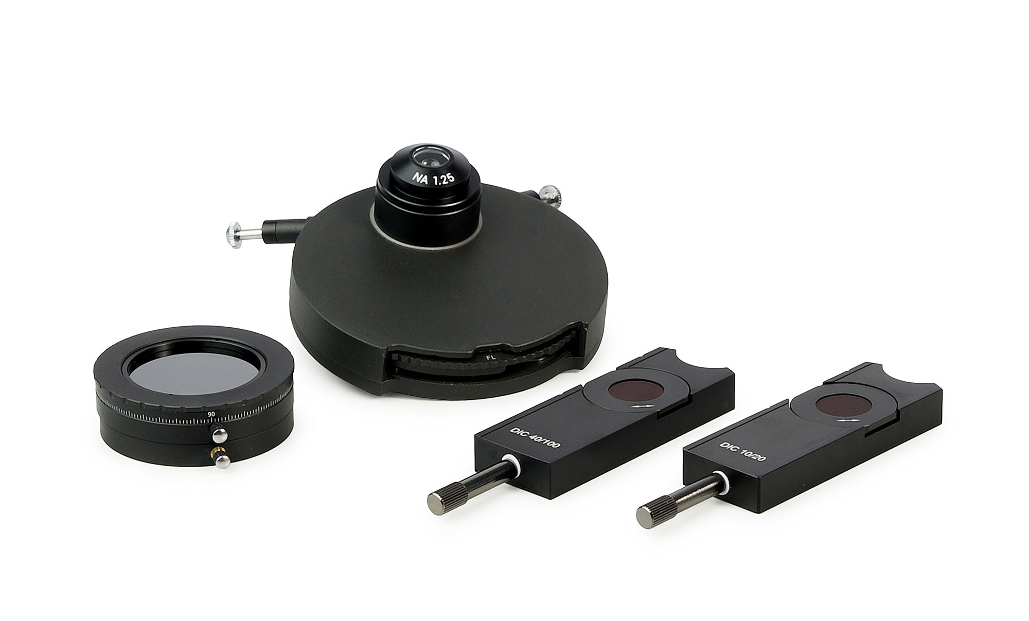
The differential-interference contrast device includes a DIC condenser polarization filter and DIC sliders.
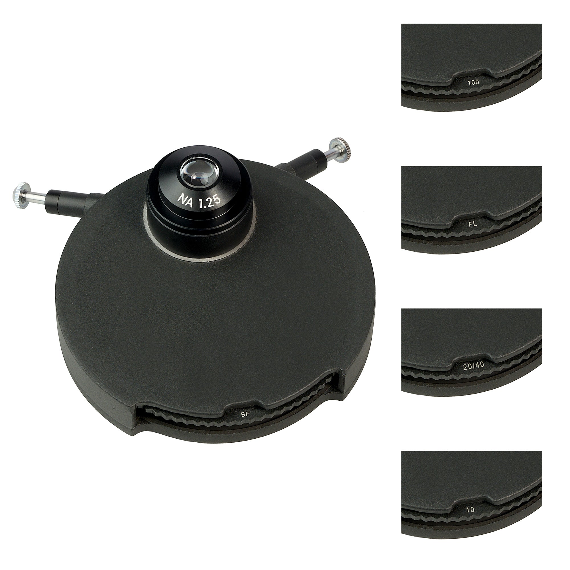
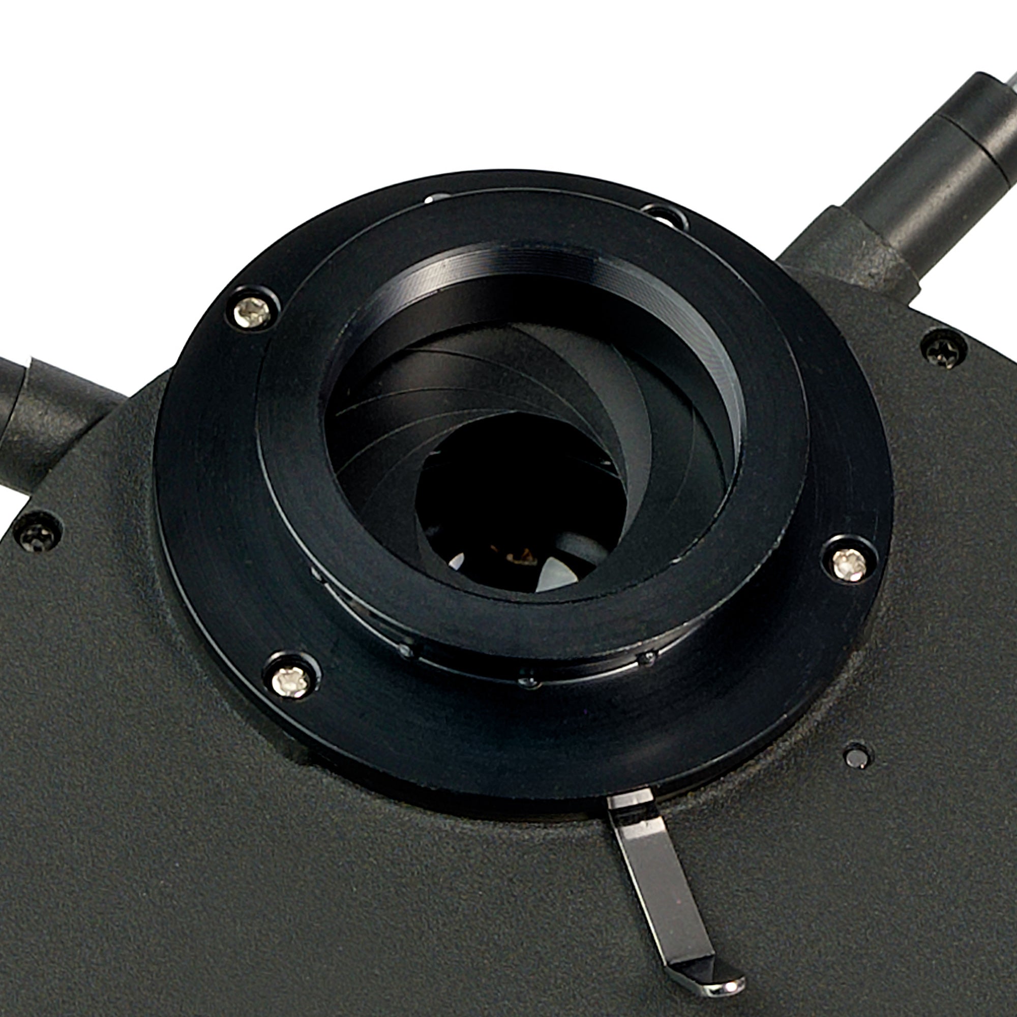
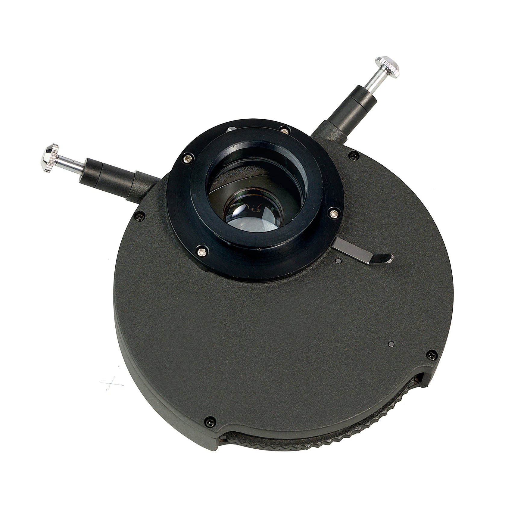
DIC condenser
The DIC condenser is installed in place of the brightfield condenser. The turret includes DIC prisms and a slot that is used during brightfield observations. The DIC prisms in the turret are used depending on the objective being used.
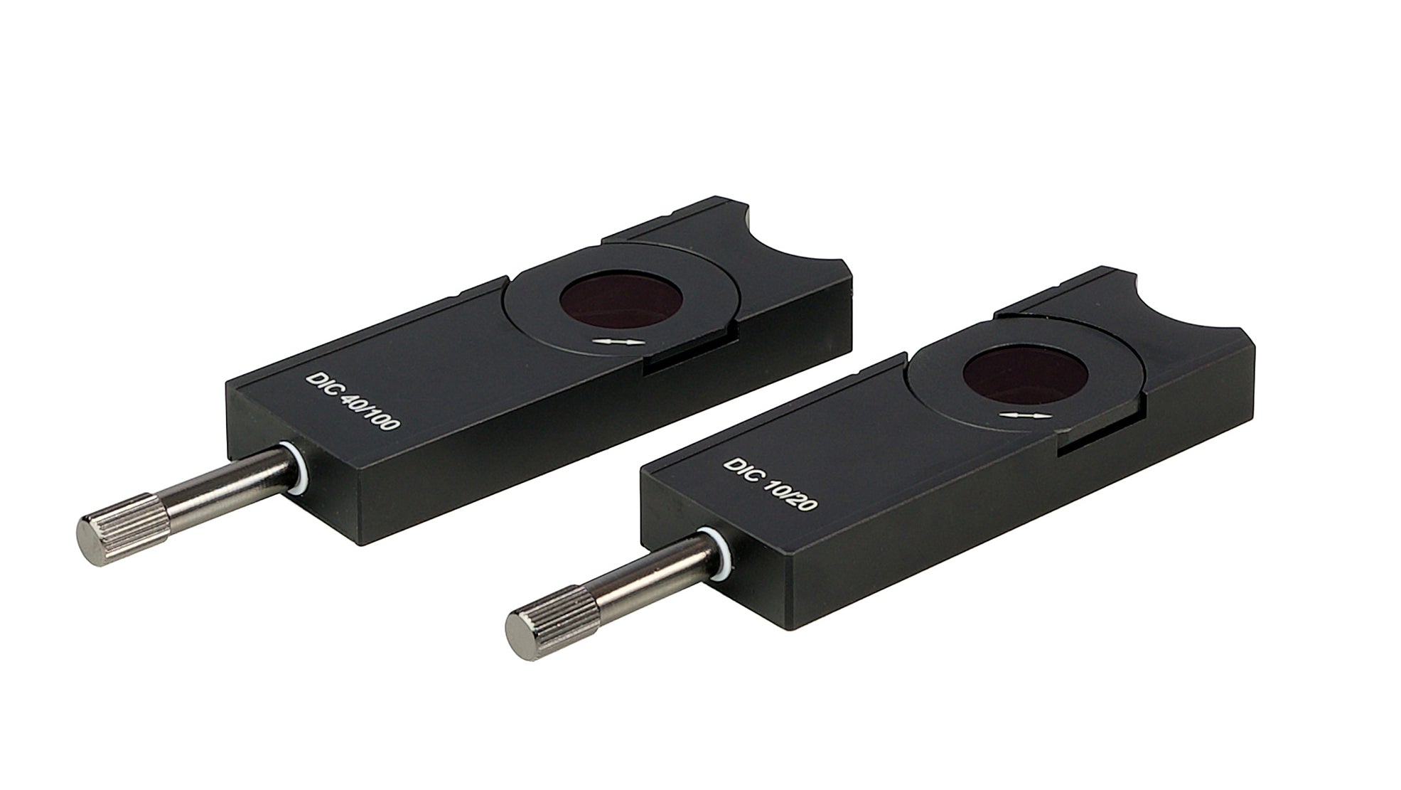
DIC sliders
The DIC slider is installed in a special slot above the revolving nosepiece. The DIC slider is used in combination with the corresponding objective. The slider is a plate with a DIC prism and an analyzer. The MAGUS DIC290 device includes two sliders: One is used with 10x and 20x objectives and the other one with 40x and 100x objectives. The rotation of the slider knob moves the DIC prism in the transverse direction.
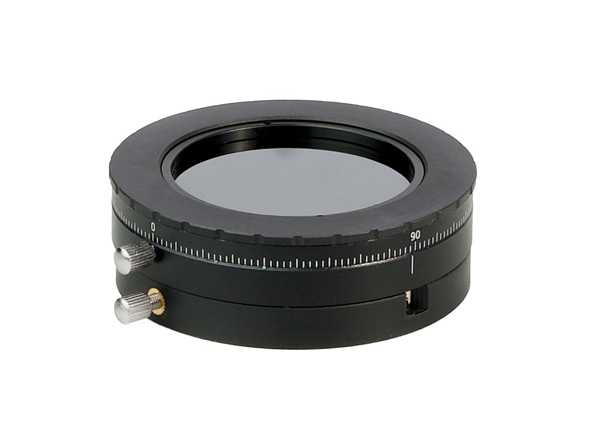
Polarization filter
When the DIC technique is used the light from the illuminator is passed through a polarization filter. The polarizer is placed on the collector.
Recommended objectives
The objectives are not included in the kit of the MAGUS DIC290 device. The DIC technique requires a precise position of the optical components relative to each other: the lower DIC prism – relative to the front focal plane of the condenser and the upper DIC prism – relative to the rear focal plane of the objective. The MAGUS DIC290 device is designed for MAGUS Bio 290T microscope and therefore you should use the microscope’s supplied or compatible objectives for DIC observations. We recommend using MAGUS FL S-APO60 PlanF objectives with a higher aperture. A higher aperture of an objective allows focusing on a thin flat section of a thick specimen which is called optical sectioning. Higher apertures also increase the resolving power of the optical microscope.
Compatibility:
Package:
| Brand | MAGUS |
| Warranty | 2 years |
| EAN | 5905555025629 |
| Package size (LxWxH) | 4x7x4 cm |
Temos uma política de devolução de 14 dias. Se não estiver satisfeito com a sua compra, devolva-a na sua embalagem original e certifique-se de que coloca a embalagem original do produto dentro da caixa de envio para evitar quaisquer danos.
Todos os artigos devem estar no seu estado original e não devem ser utilizados. O valor da sua compra será reembolsado. .
As despesas de envio não serão reembolsadas.
Se a devolução se dever a um produto defeituoso, poderá ser compensado pelo envio da devolução.
Se necessitar de efetuar uma devolução, contacte-nos para obter mais informações.
- De 0 a 2 kg - custo de envio: 12,50 euros.
- De 2.001 kg a 30 kg - custo de envio: 17,00 euros.
- A partir de 30,001 kg - custo de envio: 30,00 euros.
Em caso de recolha no nosso armazém ou pontos de revenda - o envio é gratuito.
Obrigado por subscrever!
Este e-mail foi registado!